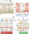
The trinity of COVID-19: immunity, inflammation and intervention
- PMID: 32346093
- PMCID: PMC7187672
- DOI: 10.1038/s41577-020-0311-8
The trinity of COVID-19: immunity, inflammation and intervention
Abstract
Severe acute respiratory syndrome coronavirus 2 (SARS-CoV-2) is the causative agent of the ongoing coronavirus disease 2019 (COVID-19) pandemic. Alongside investigations into the virology of SARS-CoV-2, understanding the fundamental physiological and immunological processes underlying the clinical manifestations of COVID-19 is vital for the identification and rational design of effective therapies. Here, we provide an overview of the pathophysiology of SARS-CoV-2 infection. We describe the interaction of SARS-CoV-2 with the immune system and the subsequent contribution of dysfunctional immune responses to disease progression. From nascent reports describing SARS-CoV-2, we make inferences on the basis of the parallel pathophysiological and immunological features of the other human coronaviruses targeting the lower respiratory tract - severe acute respiratory syndrome coronavirus (SARS-CoV) and Middle East respiratory syndrome coronavirus (MERS-CoV). Finally, we highlight the implications of these approaches for potential therapeutic interventions that target viral infection and/or immunoregulation.
Conflict of interest statement
The authors declare no competing interests.
Figures


Similar articles
- A Comprehensive Review of Animal Models for Coronaviruses: SARS-CoV-2, SARS-CoV, and MERS-CoV.Virol Sin. 2020 Jun;35(3):290-304. doi: 10.1007/s12250-020-00252-z. Epub 2020 Jun 30.Virol Sin. 2020.PMID: 32607866Free PMC article.Review.
- Highlight of Immune Pathogenic Response and Hematopathologic Effect in SARS-CoV, MERS-CoV, and SARS-Cov-2 Infection.Front Immunol. 2020 May 12;11:1022. doi: 10.3389/fimmu.2020.01022. eCollection 2020.Front Immunol. 2020.PMID: 32574260Free PMC article.Review.
- Angiotensin-converting enzyme 2 (ACE2), SARS-CoV-2 and the pathophysiology of coronavirus disease 2019 (COVID-19).J Pathol. 2020 Jul;251(3):228-248. doi: 10.1002/path.5471. Epub 2020 Jun 10.J Pathol. 2020.PMID: 32418199Free PMC article.Review.
- Tropism, replication competence, and innate immune responses of the coronavirus SARS-CoV-2 in human respiratory tract and conjunctiva: an analysis in ex-vivo and in-vitro cultures.Lancet Respir Med. 2020 Jul;8(7):687-695. doi: 10.1016/S2213-2600(20)30193-4. Epub 2020 May 7.Lancet Respir Med. 2020.PMID: 32386571Free PMC article.
- The Immune Response and Immunopathology of COVID-19.Front Immunol. 2020 Aug 26;11:2037. doi: 10.3389/fimmu.2020.02037. eCollection 2020.Front Immunol. 2020.PMID: 32983152Free PMC article.Review.
Cited by
- Thymidine Phosphorylase Is Increased in COVID-19 Patients in an Acuity-Dependent Manner.Front Med (Lausanne). 2021 Mar 22;8:653773. doi: 10.3389/fmed.2021.653773. eCollection 2021.Front Med (Lausanne). 2021.PMID: 33829029Free PMC article.
- Future Biomarkers for Infection and Inflammation in Febrile Children.Front Immunol. 2021 May 17;12:631308. doi: 10.3389/fimmu.2021.631308. eCollection 2021.Front Immunol. 2021.PMID: 34079538Free PMC article.Review.
- Difference in mortality rates in hospitalized COVID-19 patients identified by cytokine profile clustering using a machine learning approach: An outcome prediction alternative.Front Med (Lausanne). 2022 Sep 20;9:987182. doi: 10.3389/fmed.2022.987182. eCollection 2022.Front Med (Lausanne). 2022.PMID: 36203752Free PMC article.
- Pathophysiology and Potential Therapeutic Candidates for COVID-19: A Poorly Understood Arena.Front Pharmacol. 2020 Sep 17;11:585888. doi: 10.3389/fphar.2020.585888. eCollection 2020.Front Pharmacol. 2020.PMID: 33041830Free PMC article.Review.
- Integrative omics provide biological and clinical insights into acute respiratory distress syndrome.Intensive Care Med. 2021 Jul;47(7):761-771. doi: 10.1007/s00134-021-06410-5. Epub 2021 May 25.Intensive Care Med. 2021.PMID: 34032881Free PMC article.
References
- World Health Organization. WHO Director-General’s opening remarks at the media briefing on COVID-19 - 11 March 2020. WHOhttps://www.who.int/dg/speeches/detail/who-director-general-s-opening-re... (2020).
Publication types
MeSH terms
LinkOut - more resources
Full Text Sources
Other Literature Sources
Molecular Biology Databases
Miscellaneous

